



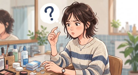
Contact lens deposits and contamination occur when tear film organic components adsorb onto the lens polymer, combined with the adhesion of microorganisms and external contaminants from fingers or the environment2, 3, 5. Soft contact lenses are exposed to a complex biochemical ocular environment immediately upon insertion; tear proteins adhere to the lens matrix within less than one minute3. The accumulation and chemical degradation of these deposits induce contact lens discomfort (CLD), disrupt tear film stability, and increase the risk of corneal epithelial damage, bacterial biofilm formation, and vision-threatening infections like microbial keratitis2, 4, 5.
Fundamental Mechanisms of Contact Lens Deposit Formation and Tear Protein Behavior
Tear film proteins are the primary component of contact lens deposits, and their adsorption kinetics are dictated by the electrostatic interactions between the electrical charges of the lens polymer and the proteins.
Upon lens insertion, over 400 distinct proteins in the tear film (ranging from 10 to 2,360 kDa) begin adhering to the biomaterial surface3. The primary tear proteins of interest in clinical contact lens research are lysozyme (14.3 kDa, isoelectric point [pI] pH 11.4), lactoferrin (80 kDa, pI pH 8.7), and albumin (66 kDa, pI pH 5.2)3.
Since the average physiological pH of the human tear film is 7.4, these proteins exhibit different net charges based on their respective isoelectric points:
・Lysozyme and Lactoferrin: Positively charged at pH 7.433.
・Albumin: Negatively charged at pH 7.433.
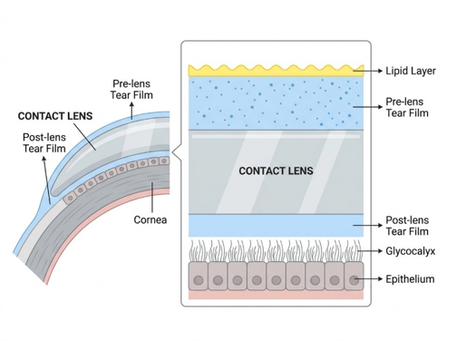
These electrical charge differentials determine how proteins interact with various lens materials:
・High Water Content Ionic Materials (FDA Group IV):
Materials such as Etafilcon A copolymerize negatively charged monomers like methacrylic acid (MAA), rendering the lens surface highly anionic3. This negative surface charge exerts a powerful electrostatic attraction on positively charged lysozyme, resulting in massive protein adsorption (averaging ~1,300 µg/lens)3. This is more than ten times the level of protein deposits typically observed on non-ionic hydrogels or other pHEMA-based materials (which generally accumulate less than 100 µg/lens)3.
・Silicone Hydrogel (SiHy) Materials:
Modern silicone hydrogel lenses generally attract far less total protein than conventional hydrogels, with non-ionic SiHy materials depositing less than 10 µg/lens and ionic SiHy materials (such as Balafilcon A) accumulating up to 34 µg/lens3. However, the relative percentage of denatured (unfolded) protein is significantly higher on silicone hydrogel lenses than on conventional hydrogel materials3.
Protein denaturation occurs when the adsorbed protein undergoes a conformational structural change (coiling randomly), losing its native biological and antimicrobial activity3. These denatured proteins are recognized by the ocular immune system as foreign, triggering inflammatory complications such as giant papillary conjunctivitis (GPC) and driving contact lens-induced discomfort and allergic ocular surface reactions2, 3.
Lipid Adsorption and Oxidative Degradation on Silicone Hydrogel Lenses
Silicone hydrogel materials readily adsorb tear film lipids due to their hydrophobic properties, and the subsequent oxidation of unsaturated fatty acids under ocular oxidative stress directly contributes to contact lens discomfort.
To achieve high oxygen transmissibility (Dk/t), silicone hydrogels incorporate siloxane macromers3. However, these silicone moieties are naturally hydrophobic and highly lipophilic, making them highly prone to attracting tear film lipids2, 3.
A clinical trial conducted by Toho University Faculty of Medicine (Itokawa et al., 2026) evaluated 38 habitual soft contact lens wearers utilizing comfilcon A silicone hydrogel lenses over a 2-week period4. By separating the participants into a symptomatic group (J-CLDEQ-8 score ≥ 11, n=25) and an asymptomatic group (J-CLDEQ-8 score < 11, n=13), and analyzing the lenses using gas chromatography-mass spectrometry (GC-MS), the researchers demonstrated a direct link between lipid chemical degradation (oxidation) and contact lens discomfort4.
Unsaturated Fatty Acid Oxidation and Subjective Discomfort
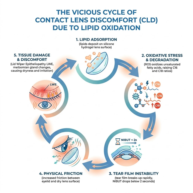
Tear film lipids (cholesteryl esters, wax esters, and phospholipids) contain various fatty acids, with C16 and C18 species being the most abundant4. Under the constant oxidative stress and inflammatory conditions present on the ocular surface, "unsaturated fatty acids" (which contain double bonds, such as palmitoleic acid C16:1 and oleic acid C18:1) are highly susceptible to lipid peroxidation driven by reactive oxygen species (ROS)4.
This degradation process generates highly reactive peroxides and short-chain aldehydes (such as malondialdehyde [MDA], nonanal, and heptanal), which cause chemical irritation to the corneal and conjunctival epithelia, significantly worsening dry eye and discomfort symptoms4.
As unsaturated fatty acids degrade, the ratio of stable "saturated fatty acids" to their unsaturated counterparts of the same carbon number increases on the lens4. According to the clinical data from Itokawa et al. (2026), these ratios were significantly higher in the symptomatic group4:
・Palmitic Acid (C16:0) / Palmitoleic Acid (C16:1) Ratio:
Symptomatic wearers exhibited a ratio of 4.73 ± 3.51, whereas asymptomatic wearers showed 2.13 ± 1.10—representing a statistically significant ~2.2-fold increase (p = 0.0219)4.
・Stearic Acid (C18:0) / Oleic Acid (C18:1) Ratio:
Symptomatic wearers showed a ratio of 3.64 ± 3.15, compared to 1.06 ± 0.76 in the asymptomatic group—a statistically significant ~3.4-fold increase (p = 0.0415)4.
These fatty acid ratios demonstrated a powerful positive correlation with the J-CLDEQ-8 questionnaire scores (r = 0.64, p < 0.001 for C16; r = 0.57, p < 0.01 for C18), validating them as robust biochemical biomarkers for evaluating contact lens discomfort4.
Tear Film Instability and Friction-Induced Tissue Damage
When fatty acids oxidize, the structural integrity of the thin tear film lipid layer (TFLL, 50–100 nm thick) collapses, causing a loss of its primary function: retarding aqueous tear evaporation2.
In the Toho University study, symptomatic wearers demonstrated a significantly shorter non-invasive tear break-up time (NIBUT) after 2 weeks of lens wear (1.8 ± 2.1 seconds) compared to the asymptomatic group (3.8 ± 2.9 seconds, p = 0.0141)4. This rapid tear film collapse severely reduces lens surface wettability, exponentially increasing physical friction between the palpebral conjunctiva (inner eyelid) and the lens surface2, 4.
This chronic mechanical friction triggers physical tissue alterations4:
・Lid Wiper Epitheliopathy (LWE):
Symptomatic wearers showed significant worsening of upper eyelid LWE grades over 2 weeks (progressing from 1.2 ± 0.6 at 15 minutes to 1.5 ± 0.6 at 2 weeks, p = 0.0369)4.
・Meibomian Gland Dysfunction (MGD):
Upper eyelid meibomian gland loss (meiboscore) was significantly worse in the symptomatic group (1.5 ± 0.8) than in the asymptomatic group (1.0 ± 0.5, p = 0.0222), locking the patient into a chronic spiral of dry eye and discomfort4.
Importantly, the study noted that healthy, non-oxidized squalene (a unique skin/tear lipid) has a highly beneficial effect on silicone hydrogel lenses4. Squalene deposition significantly correlates with improved tear film stability (r = 0.4060, p = 0.0190) by reducing water evaporation4. Thus, preventing the chemical "oxidation/degradation of unsaturated fatty acids" while preserving healthy lipid spread is the critical pathway to maintaining long-term ocular comfort4.
Scientific Evidence on Contact Lens Contamination and Bacterial Biofilms
Inadequate contact lens care and improper lens case handling facilitate the development of resilient bacterial biofilms, significantly multiplying the risk of ocular infections and inflammatory conditions.
Because contact lenses physically compartmentalize the tear film and compromise the eye's natural anatomical defenses, proper hygiene is essential to prevent ocular pathology2. Epidemiological data shows that severe infectious microbial keratitis (MK) occurs in approximately 2 per 10,000 daily wearers annually, while non-infectious ocular surface inflammation (such as CLARE or CLPU) is much more common, affecting 2 to 6 per 100 wearers annually5.
Common Pathogens and Biofilm Formation
The most frequently isolated pathogen in contact lens-related microbial keratitis worldwide is Pseudomonas aeruginosa, accounting for over 70% of confirmed cases5. Gram-positive Staphylococcus aureus is also a major causative agent5.
When these bacteria colonize the surface of a contact lens or the inner walls of a lens case, they secrete an extracellular polymeric matrix, forming a highly organized biofilm5. Within a biofilm, bacteria are protected from host immune cells, ophthalmic antibiotics, and standard multipurpose disinfecting solutions (MPDS), making them extremely difficult to eradicate or wash away through simple liquid rinsing alone5.
Structured Clinical and Research Data
To provide clear, objective, and structured evidence regarding clinical parameters and hygiene regimens, key data from major third-party clinical trials are detailed in the markdown tables below.
Table 1: Comparison of Clinical Parameters in Asymptomatic vs. Symptomatic Wearers After 2 Weeks (Source: Itokawa et al., 2026; comfilcon A lenses worn for 2 weeks4)
|
Clinical Parameter |
Asymptomatic Group (n=13) |
Symptomatic Group (n=25) |
p-value |
Clinical Significance |
|
J-CLDEQ-8 Score (points) |
6.5 ± 2.7 |
17.8 ± 6.4 |
<0.0001 |
Scores ≥11 indicate symptomatic contact lens discomfort (CLD). |
|
Dryness Score (VAS, mm) |
9.4 ± 14.9 |
45.3 ± 27.5 |
<0.0001 |
Symptomatic wearers experienced substantially greater dryness. |
|
NIBUT (s) |
3.8 ± 2.9 |
1.8 ± 2.1 |
0.0141 |
Approximately 2.1-fold shorter tear film stability in symptomatic wearers. |
|
C16 Fatty Acid Ratio (C16:0/C16:1) |
2.13 ± 1.10 |
4.73 ± 3.51 |
0.0219 |
Approximately 2.2-fold increase, suggesting palmitoleic acid oxidation. |
|
C18 Fatty Acid Ratio (C18:0/C18:1) |
1.06 ± 0.76 |
3.64 ± 3.15 |
0.0415 |
Approximately 3.4-fold increase, suggesting oleic acid oxidation. |
|
Meiboscore (Upper Eyelid) |
1.0 ± 0.5 |
1.5 ± 0.8 |
0.0222 |
Greater meibomian gland loss in symptomatic wearers. |
|
LWE Grade (Upper Eyelid) |
1.7 ± 0.8 |
1.5 ± 0.6 |
0.0369 |
Symptomatic wearers showed significant worsening from baseline over the 2-week period. |
Table 2: Efficacy of Lens Case Care Regimens on Bacterial Biofilm Reduction (Source: Willcox et al., 2023; UNSW Sydney Clinical Trial5)
|
Lens Case Hygiene Protocol |
Median Bacterial Count (Range) |
Bacterial Reduction Efficacy |
p-value |
Clinical Significance |
|
Standard Manufacturer Guide |
28 cfu (0-100,000) |
Reference |
Reference |
Rinsing alone is insufficient to physically remove established bacterial biofilms. |
|
Recommended Wiping Protocol |
12 cfu (0-10,000) |
Approximately 50-60% reduction in contamination (≈0.4×) |
P=0.004 |
Strongly recommended. Physical wiping disrupts bacterial biofilms and significantly reduces contamination. |
Recommended Preventive Daily Habits and Lens Care Practices
Meticulous adherence to physical rubbing, tissue wiping, proper drying, and adopting hygienic lens packaging design concepts can dramatically prevent lens deposits and bacterial contamination.
By implementing evidence-based daily habits, contact lens wearers can actively protect their ocular surface health and optimize wearing comfort.
1. Advanced Lens Case Care (The Wiping and Air-Drying Habit)
According to epidemiological research by Stapleton et al. at UNSW, performing two simple habits—"replacing the lens case at least every 3 months" and "completely air-drying the case face down"—reduces the overall risk of microbial keratitis by 62% (nearly two-thirds)5.
Specifically, air-drying alone decreases risk by 49%, and quarterly replacement reduces it by 27%5. Furthermore, as demonstrated in Table 2, adding a physical "wiping step with a clean dry tissue" to the case care routine removes adherent bacterial biofilms, cutting the bacterial load in half5.
2. Manual "Rub and Rinse" of the Lenses
For frequent replacement soft contact lenses (2-week or monthly schedules), a daily manual "rub and rinse" is indispensable5.
After removing the lens from the eye with clean, dry fingers, the user should place the lens in the palm of their hand, apply several drops of fresh disinfecting solution, and gently rub both surfaces of the lens with a finger pad5. This physical friction is required to break the electrostatic bonds of denatured proteins and remove oxidized lipids3. Neglecting this step causes denatured lysozyme and rancid fatty acids to permanently bind to the polymer, leading to chronic ocular surface friction and discomfort3, 5.
3. Improving Blinking Habits During Screen Use
When staring at digital screens (computers, tablets, smartphones), the human blink rate drops drastically from a baseline of ~15 blinks per minute down to ~8 blinks during reading, and ~4 blinks during active screen use2.
This severe reduction is accompanied by a higher rate of "partial blinking" (incomplete blinks where the eyelid fails to fully cover the cornea)2. Incomplete blinking extends the exposed interblink interval (IBI) to 10–30 seconds, causing rapid local evaporation of the tear film and forming dry spots on the lens surface2. This localized dewetting accelerates lipid binding and permanent deposition2. Consciously practicing complete, deep blinking during digital device use acts as a vital physical defense mechanism for the pre-lens tear film2.
4. Hygienic Packaging Innovations (Touch-Free Concepts)
A critical pathway to avoiding early lens contamination is minimizing physical contact with the lens during the insertion process2, 5.
Traditional domed contact lens blisters require the user to reach into the packaging, inevitably touching the inner (concave, cornea-facing) surface of the lens with their finger2, 5. Even with hand washing, this finger contact transfers residual skin lipids (sebum, fatty acids) and epidermal microbes directly onto the lens inner surface2, 5.
Adopting innovative "smart packaging designs" that ensure the lens is always oriented with its outer surface facing upward allows the user to pinch and insert the lens without ever touching the cornea-facing inner surface2, 5. Preventing this initial sebum and microbial transfer is clinically shown to preserve pre-lens tear film stability, prevent early lipid degradation, and significantly reduce the risk of contact lens discomfort and infectious complications2, 4.
FAQ
Q. I rinse my lens case with disinfectant and dry it every day. Why is there still a risk of bacterial contamination?
A. Bacteria such as Pseudomonas aeruginosa form resilient "biofilms" on case walls by secreting a protective polysaccharide matrix5. This matrix shields them from chemical disinfectants and cannot be removed by simple liquid rinsing5. Incorporating a physical "wiping step" with a clean dry tissue after rubbing and rinsing breaks down the biofilm structure and reduces bacterial levels by approximately half5.
Q. Does using computers or screens for long hours really cause contact lenses to get dirtier?
A. Yes. When focusing on digital screens, the human blink rate decreases by nearly 75% (from ~15 to ~4 blinks per minute)2. This is combined with a high rate of incomplete or partial blinks2. The resulting dry intervals (lasting 10–30 seconds) allow the tear film over the lens to collapse, facilitating the rapid binding and accumulation of stubborn lipid deposits2.
Q. Silicone hydrogel lenses are advertised as advanced. Why do they still cause dryness and discomfort?
A. While silicone hydrogels are highly resistant to protein deposits, their silicone components are hydrophobic and lipophilic3. This means they attract tear film lipids far more easily than traditional hydrogels2, 3. These lipids (specifically unsaturated fatty acids) oxidize under the ocular surface's oxidative stress, generating chemical irritants that trigger dryness and contact lens discomfort4.
Key Takeaways
- Deposits Form Instantly: Ocular proteins and lipids begin adsorbing to contact lens surfaces within less than one minute of lens insertion3 .
- Unsaturated Lipid Oxidation Drives CLD: The chemical oxidation of unsaturated fatty acids on silicone hydrogel lenses increases the saturated/unsaturated fatty acid ratios by 2.2 to 3.4-fold, generating irritating chemical byproducts that directly cause contact lens discomfort4 .
- The Tear Collapse-Friction Spiral: Worn lenses with unstable tear films (NIBUT < 2 seconds) experience severe mechanical friction, resulting in upper eyelid tissue damage (LWE) and meibomian gland loss4.
- Wiping Cases Destroys Biofilm: Adding a physical "tissue wiping" step to lens case cleaning cuts the median bacterial contamination level in half compared to passive disinfectant rinsing5 .
- Smart Packaging Concept Blocks Sebum Transfer: Lens packaging designs that prevent fingers from touching the lens's inner (cornea-facing) surface effectively eliminate early lipid contamination and microbial transfer2, 5 .
References
-
Morgan PB, Woods C, Tranoudis IG, Helland M, Efron N, Knajian R, et al. International contact lens prescribing in 2009. Contact Lens Spectrum. 2010;25:30-53.
-
Craig JP, Willcox MDP, Argüeso P, Maissa C, Stahl U, Tomlinson A, et al. The TFOS International Workshop on Contact Lens Discomfort: Report of the Contact Lens Interactions With the Tear Film Subcommittee. Invest Ophthalmol Vis Sci. 2013;54(11):TFOS123-TFOS156.
-
Luensmann D, Jones L. Protein deposition on contact lenses: The past, the present, and the future. Contact Lens Anterior Eye. 2012;35(2):53-64.
- Itokawa T, Suzuki T, Kakisu K, Hori Y. Impact of lipid degradation and tear film stability on silicone hydrogel contact lens wearers with discomfort. Contact Lens Anterior Eye. 2026;49(2):102616 (doi:10.1016/j.clae.2026.102616).
- Willcox MDP, Bahatheg G, Carnt N, Kalaiselvan P, Kumar N, Kuppusamy R, et al. Biofilms and contact lenses: problems and solutions. Microbiology Australia. 2023;44(2):96-99.
Reviewed by: Menicon Clinical Affairs
Updated: September 28, 2026